Introduction
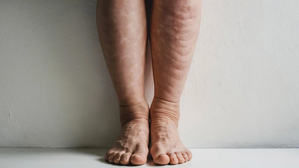
Lymphedema is a chronic and a progressive condition which results in accumulation of protein rich fluids in the interstitium. This leads to amplified inflammatory response and deposition of fat and fibrous tissue. Lymphedema begins with pain, discomfort and pitting edema, progressing to fat deposition and permanent disfigurement. These patients get repeated attacks of cellulitis and need long term antibiotics. Increased limb size can interfere with mobility and affect body image leading to significant physical and psychological morbidity. These patients are usually untreated, unaware and misdirected with respect to the right treating speciality.
Most common cause of lymphedema worldwide and developing countries is still infective due to Filariasis. However in developed countries, lymphedema due to post oncological resection remans the leading cause. Incidence of lymphedema depends upon on how extensive the disease and resection is. The incidence of lymphedema after breast cancer treatment ranges from 24% to 49% after mastectomy and 4% to 28% after lumpectomy. Even less invasive procedures like sentinel node biopsy is associated with only a 5% to 7% incidence of upper extremity lymphedema.
Traditionally these patients have been ignored and misdirected. Only a fraction of these patients end up receiving standard physiotherapy protocol. Several recent systematic reviews have highlighted the distinct lack of evidence for the optimal management of lymphedema. Physiotherapy has been the only effective treatment modality so far. However, it is cumbersome, time consuming and a costly affair. There has been a paradigm shift in management of lymphedema with the advent of microsurgical procedures. Vascularised lymph node transfers and lympho-venous anastomosis have been added in the armamentarium and plastic surgeons play a vital role in managing these patients.
Despite advances in microsurgery, there is neither consensus on surgical or nonsurgical procedures nor a standardized protocol in the treatment of the patient with lymphedema.
Non-surgical management
Treatment of lymphedema include both non-surgical and surgical management. Non-surgical management is a pre-requisite in all cases.
Goal of non-surgical lymphedema management is :
- Exercise and Joint moility
- Swelling reduction and maintenance
- Skin care
- Risk reduction
- Pain and Psychological management
This involves combination of compression and exercise with or without lymphatic massage (manual lymphatic drainage [MLD] or intermittent pneumatic compression [IPC]).
Manual lymphatic Drainage (MLD)
Aims are increasing the drainage of the lymph through the remaining patent lymphatic channels, avoiding the ineffective and obliterated vessels. MLD is advocated by most as the primary treatment modality. However, currently various randomised control trials suggest little evidence supporting the use of massage alone.
Furthermore, aggressively performed MLD can be counterproductive and can lead to increased oedema due to damage of lymphatic vessels and capillaries.
Combined physical therapy and Compressive therapy
Combined physical therapy (CPT), also known as complete or complex decongestive therapy (CDT), is also a two-stage treatment protocol.
In phase I, the goals are limb size reduction and improvement of the skin quality. Proceeding to phase II, the aim is to consolidate the gains of phase I in an individualised manner. There is a long standing evidence on the efficacy of CPT in managing lymphedema. Although a labour intensive procedure, it is considered a standard of care. Compliance is imperative for successful outcomes
Phase 1
- Skin care
- MLD
- Range of motion exercises
- Multi-layered bandaging.
Multilayer bandage wrapping is the mainstay of conservative therapy.
Phase 2 – Maintaining the results of phase I with the use of a low-stretch elastic stocking or sleeve compression.
With CPT, randomized, controlled studies have shown a mean decrease of between 40% and 60% in edema volume. Compressive bandages can act like a constriction band across the joints if not adequately padded. This can lead to increased lymphedema in the distal limb. Hence a non-individualised bandaging by an untrained person can be more harmful.
Medications
There is extensive data on the usage of drugs in lymphedema. Diuretics have been used in the first stage of lymphedema, however leads to significant fluid and electrolyte imbalance without any significant benefit in reducing peripheral edema. The only role of antibiotics in lymphedema is restricted to control cellulitis and lymphangitis. Diethylcarbamazine, albendazole, or ivermectin have been used for controlling the active phase of filariasis only.
Intermittent Pneumatic Compression (IPC)
It is an external compression sleeve with multiple air chambers which create a gradient compression from the distal limb to the proximal nodal basin. More than augmenting lymphatic drainage, IPC reduces the capillary filtration. IPC is a controversial treatment modality, which can harm the superficial lymphatics with high pressure. Also, contraindicated in patients with renal, cardiac anomalies and those with lower limb malignancies.
Exercise and elevation
Individualised as per patient requirement, gentle exercises, mobility training and aerobics help to improve lymphatic drainage through the muscular pump. Has an impact on the overall psychological and functional well-being of the individual. Limb elevation above the heart level helps in improving the edema by increasing the venous drainage.
Diet and weight loss
In an already obese individual weight loss has been proven to be essential in controlling lymphedema. However, just weight reduction cannot cure the pathology. No dietary modification has been proven to be of any use in controlling lymphedema. Fluid restriction has no role either.
Skin care
Skin hygiene and care are vital to the success of virtually all treatment approaches. Skin is the primary portal of entry of bacterial and fungal pathogens which lead to repeated attacks of cellulitis. Each attach of cellulitis (Acute Dermato-lymphangio-adenitis – ADLA) further worsens lymphedema and affect the skin quality. Maintaining the skin barrier is crucial for the long term outcome in progress of lymphedema. pH neutral soaps with glycerine are preferrable for cleansing, avoid irritant and allergic products. Emollients are crucial in creating a lipid layer over the skin hence preventing water loss and control microbial entry.
Surgical Management
Liposuction
Liposuction is offered as a reduction procedure to patients with non-pitting edema without skin changes. This subset of patients have predominantly fat dominant lymphedema. Physiological surgeries in these patients do not achieve significant reduction in limb size as adipose tissue deposition cannot be reversed.
Pre-operative MRI will help to quantify and plan the extent of liposuction. Brosnon has standardised the technique of liposuction. He advocates use of power-assisted liposuction using 3mm cannula size through multiple entry points under tourniquet control. Customized pressure garments are designed pre-operatively and is applied on table under anesthesia. Results are ideally observed at 3-6 months from surgery and need to continue wearing customised pressure garments.
Excisional Procedure
Excisional procedures are reserved for patients with severe fibrotic changes and limb deformity. These patients are typically stage 3 (ISL classification) with long term lymphedema, non-compliant to physiotherapy and repeated attacks of cellulitis.
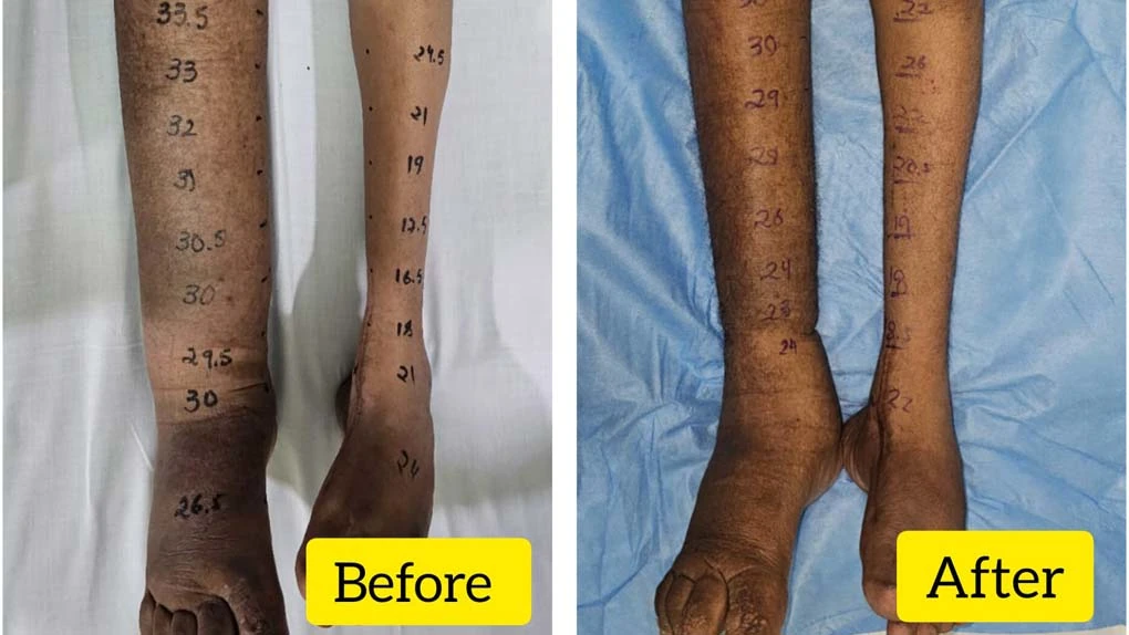
The lymphatic channels in these patients are damaged and very little can be expected out of physiological surgeries. Charles excision is the most popular technique among various reduction surgeries that have been documented. Traditionally was described in patients with filarial lymphedema involving the scrotum. The principles of the excision are now extended to lower limb as well. This involves circumferential excision of the skin and subcutaneous tissue, leaving behind the deep fascia. The raw area is grafting. There is high complication rate of recurrence, graft loss, non-healing ulcers and infections which is amplified with larger area. There are reports of combining Charles excision with vascularised lymph-node transfer as well with encouraging results a fewer long term complications. The goal of these surgeries is to have improved skin hygiene, reduced functional morbidity, risk of cellulitis and application of pressure garments. Karri et al reported subjective improvements in mobility with acceptable cosmetic results and low complication rates. Salgado et al, is the only one who has reported outcomes of volume reduction as a percentage reduction in Charles excision.
Vascularised lymph node transfer
Vascularised lymph node transfer (VLNT) is a physiological surgery which improves the lymphatic drainage of the limb. Local release of vascular endothelial growth factor 3 and stimulates lymphangiogenesis, hence acting as a sponge or a wick to absorb the accumulated interstitial lymph. It absorbs the lymphatic fluid and diverts it into the venous track. Physical therapy can improve the drainage to a particular point only and there after physiological procedures can only lead to further improvement and stabilization. Ideally described for patients with secondary lymphedema and hypoplastic congenital lymphedema.
Multiple sources of VLNT have been described and advocated without proven superiority. Commonly used flaps are groin flap (with or without DIEP), Sub-mental flap, Supra-clavicular, lateral thoracic and Omental flap. Harvesting these flaps require a good knowledge of the anatomy and microvascular skill set. MR lymphangiography, although a challenging procedure is ideal for diagnosis and comparison post VLNT.
Corinne becker advocates the placement of the adipo-lymphoid nodal tissue in the previously operated site. Understanding of axillary anatomy and release of post-radiation scar tissue is of utmost importance to avoid damaging any vital structure. Few others advocate placement of the tissue in the most distal part of the limb, so as to take advantage of the gravity assisted pooling of lymph and the sponge like property of the VLNT. Degree of improvement from VLNT depends upon the stage of the disease, extent of fibrosis and fat deposition, quality of skin, age of patient and duration of lymphedema.
According to Corinne’s evaluation of 1500 cases, patients with stage 1 lymphedema observed a reduction of 40 % in upper limb and 33 % in lower limb volume. Few patients at the end of 2 years had complete resolution without the need of pressure garments. Those with stage 2 or 3 experienced an improvement of 60%, however were still dependent on compression garments. For better aesthetic outcome these patients required selective liposuction. In both cases there was a significant improvement in the infection rates, pain and discomfort. Earlier the diagnosis, better the result. MR lymphangiography is a far superior imaging modality that lymphoscintography to document any improvement. Demonstrates reduced dermal backflow and new lymphatic tracks in 50% of the patients. Patients with hypoplastic congenital lymphedema diagnosed on MR lymphangiography are selected for VLNT. Logically will stimulate lymph-angiogenesis and improve the drainage.
All patients required post-operative physiotherapy for 3 months and gradually weaned off. Some degree of improvement is expected in all patients. Even patients with elephantiasis can be treated with combined approach of Charles excision and VLNT. This treatment modality is advocated to reduce infection rates, recurrence and need for physical therapy.
Lymphatico-venous anastomosis
LVA is a microsurgical technique which involves anastomosis of sub-dermal lymphatics to sub-dermal venules (0.5 – 0.8 mm). The technique demands super-microsurgical skills, instrumentation and higher magnification (30 X). Koshima advocates using 50 micron needle for suturing to prevent vessel injury. The procedure can be performed under local anaesthesia and doesn’t require long term admission. There are various techniques which help to identify and visualise the lymphatics tracks. ICG dye is injected in the sub-dermal plane or in the superficial fatty layer. Lymphatics channels that might be functional are visualised using a handheld ICG camera. More often we find a track along the medial and lateral aspect of the limb and in the distal arm and leg. Under local anaesthesia a 3cm perpendicular incision is made. Iso-sulfane blue is an excellent dye to visualise lymphatic ducts and injected adjacent to the planned incision site.
Through the same incision an adjoining venule is identified and various techniques of anastomosis are described (End to End, End to Side and Octopus Technique). Octopus techniques involves anastomosing multiple lymphatic vessels into a relatively larger venule. This helps to overcome the lumen size discrepancy and preventing venous backflow into the lymphatic tracks. Few authors advocate using a 6-0 prolene to cannulate the lymphatic vessel for the ease of suturing. Theoretically for better lymphatic drainage, both antegrade and retrograde anastomosis of the lymphatic vessels to the venule is advised. There is a significant variation with the number of anastomosis performed (range 2-9; average 3) per patient and combined with various supplementary procedures like DCT or reduction surgeries. Most studies show an improvement in limb volume and significant reduction in incidence of cellulitis. Chang has reported an volume improvement of 42% and 96% improvement in subjective scoring in his series of 100 consecutive cases. Various other studies also demonstrates similar results.
In patients with severe skin fibrosis it is technically challenging to find a patent functional lymphatics. However, deeper lymphatics are usually patent and can be anastomosed to the venule.
LYMPHA
Lymphatic Microsurgical Preventive Healing Approach (LYMPHA) is a prophylactic surgery in patients undergoing axillary dissection for carcinoma breast. Approximately 25% (13 to 52%) of the patients with nodal excision and radiotherapy develop lymphedema and even 6% of patients post axillary sampling.
LYMPHA can be offered to patients which are at relatively higher risk of developing lymphedema. These patients are those with BMI greater than 30 and transport index >/= 10 on lymphangiography. A blue dye (1-2ml) is injected subdermal or sub-cutaneous on the medial arm and the nodal tissue draining the arm are identified.
After resection of these nodes the lymphatic channels are buried in a venous branch using a U stitch technique. Multiple stitches between the lymphatic adventitia and the vein wall are used to hold the anastomosis together. Commonly we find a draining node below the axillary vein and with the second costo-brachial nerve.
In Francescos, series of 74 patients undergoing LYMPHA, persistant lymphedema was seen in 3 cases only (4.05%). Transient lymphedema in 8 cases. Post-operative lymphoscintigraphy showed patent lympho-venous anastomosis and improved transport index with faster liver uptake in all patients.
Have we really found an answer ?
Looking at the current data available, we are still far away from finding the right answer or protocol in preventing and treating lymphedema. Although microsurgical procedures have helped us to move drastically in this direction, there is still no consensus. Furthermore, the philosophy and techniques of lymphedema surgery vary in different parts of the world. Filarial and congenital lymphedema are even less researched and understood, which prevents us from generalising treatment principles.
Looking at the current trends, a sound treatment plan includes a multi-modality approach, which we also follow at TMH. One year disease free interval is a must for all patients. They are started on Complex Decongested Therapy by a dedicated trained physiotherapist. Emphasising on skin care and multi-layered bandaging. Patients who present with an active episode of cellulitis are first treated with a course of antibiotics. Lympho-scintigraphy is performed pre-operatively to diagnose and have a baseline parameter for comparison. Volumetric calculations are used as the primary parameter of comparison. After counselling and detailed surgical planning, VLNT with LVA is planned based on the stage of the disease and the availability of a healthy lymphatic channels. Intra-operative ICG imaging is used to identify any patent lymphatic tracks. CDT is continued post operatively and eventually moved on to a pressure garment. Post-operative Lymphoscintigraphy is performed at 6 months and 1 year. Selective Liposuction is planned for patients 1 year post physiological surgeries for localised reduction of fatty tissue and better aesthetic outcome.